Identification of Common Worms in Saltwater Aquaria

Christmas Tree Worms

The “Identification” series of posts is to help with identifying various critters in a typical saltwater aquarium (such as this post for identification of common worms) or to just see what you may come across one day.  Marine worms come in a vast array of sizes, shapes, colors, and desirability. Most of these shown were hitchhikers on corals and live rock.  These are all creatures I’ve come across on my own (not stock photos), so there’s a good chance that you may as well. Not all marine worms are good, so it’s extremely important to quarantine or at least dip. Hopefully these photos and accompanying information will help with your identification of common worms in saltwater aquaria.

Click on an image below to read more about the worm. [Note: Sometimes the links do not immediately load, in case you’re unable to select an image. Just scroll down.]

Phylum: Annelida

Class: Polychaeta

Order: Sabellida

“Feather Dusters” and “Cocoa Worms” (Sabellidae genera/Serpulidae genera) – Reef Safe:
Marine Feather Duster colony

Appearance: Feather dusters are filter-feeding worms that live in a paper-like or cement-like tube (often encased in rock or the sand.)  The “feathers” are called radioles, and cilia (finger-like projections) on the radioles move captured food particles and sediment to the center.  The sediment is used to build the tube. 

Behavior: Most that come in as hitchhikers will do just fine in the home aquarium (like the ones shown encased in the liverock), but larger ones sold at stores (like the “Coco Worms” shown below) often slowly perish in all but a few aquariums (likely from a lack of food).  Two problems are frequently encountered with feather dusters:  they leave their tube or drop the crown of feathers (worm remains in the tube).  Dropping the crown may suggest unsatisfactory water conditions or starvation.  Be patient, and the worm will likely regrow the crown when conditions improve (assuming the conditions do improve quickly).  If the worm leaves the tube, do not try to shove it back in.  Just leave it alone, and it will find a new spot in most cases. Basically, if you have a feather duster hitchhiker, place it in lower flow (cilia are more efficient) and enjoy it! (And be mindful of predators, as certain fish may find these as a tasty snack.)

Taxonomy: “Feather dusters” are in the Phylum Annelida, Class Polychaeta, Order Sabellida, and Family Sabellidae (usually soft-tubed) or Family Serpulidae (usually hard-tubed).

“Cocoa Worms” are in the Phylum Annelida, Class Polychaeta, Order Sabellida, Family Serpulidae, Genus Protula.

Feather Duster in Catalina Island, CA, USA

Back to top

“Christmas Tree Worms” (Spirobranchus spp.) – Reef Safe:
Christmas Tree Worms

Appearance: Christmas Tree Worms are gorgeous and live up to their names; they’re shaped like their namesake trees and come in a rainbow of colors.

Behavior: Changes to the local environment (e.g., sudden shade/light, different water flow) will cause these worms to quickly retract, which makes them a quirky novelty in the home aquarium. Unfortunately, their desirability in the hobby does not match their resiliency; they do not typically live long in the hobbyist aquarium (although, there are cases of explosive growth. Your mileage may vary.)

Taxonomy: Christmas Tree Worms are in the Phylum Annelida, Class Polychaeta, Order Sabellida, Family Serpulidae, Genus Spirobranchus.

Back to top

“Spirorbid Worms” (Spirorbis spp.) – Reef Safe:
Spirorbid feather worms in marine aquarium

Appearance: Spirorbid worms are super tiny, up to only a couple of millimeters in diameter. Their white shells curve around into a circle and are stationary upon the substrate (glass, shown here).

Behavior: These are essentially miniature hard-tube feather dusters that typically cover substrate in low-flow areas.  They are harmless filter feeders, but they can clog pumps and other equipment if their population gets out of control. And, they can create quite hard and sharp projections that can easily scratch skin, so use caution around them while conducting maintenance!

Taxonomy: Spirorbid worms are in the Phylum Annelida, Class Polychaeta, Order Sabellida, Family Serpulidae, and Genus Spirorbis.

Back to top

Order: Polychaeta

“Bristle Worms” (Polychaeta) – Reef Safe with Caution:
Harmless Bristleworms

“Bristle Worms” from a common-name perspective also apply to “Fire Worms” and just about any other Polychaeta class worm, as most worms with the prominent bristles just naturally get the former name (think of it like the “Unidentified Flying Object” (UFO) nomenclature of the ocean. If it has bristles, it’s a “bristle worm”). However, I’m using them as two separate names here, as the reefkeeping hobby does to help distinguish risk between the two main polychaetes encountered. (Note: These orange “bristle worms” shown are not “Oenone fulgida,” which are predatory.)

I have to admit, these things terrify me (they always have and always will). My husband has had quite a few chuckles from watching me doing the “heebie-jeebie” dance every time I nearly touch one. The stings from touching one are quite painful but can typically be resolved with home care (of course, seek professional medical attention when needed).

Appearance: The bristle worms shown here are long (about 2-6 inches in length, although they can be much shorter or longer), slender, and are covered in bristles used for movement (and probably protection, as a dual-function). They’re typically orange (sometimes described as pink, but my eyes don’t see it), and some are more gray/blue.

Behavior: Regardless of their scary outer appearance, these worms are scavengers. They eat detritus and decaying matter (e.g., dying fish, fish excrement, leftover fish food). If you see a bristle worm on a dead fish, the fish was likely dead when the worm came along (these rarely kill fish). However, some of these can become insanely huge and predatory. Removal is best for extremely large worms (i.e., over six inches long), such as the one in the photo below. Typically these larger bristle worms have more of a gray and dull orange hue, rather than the bright orange of the smaller ones.

Taxonomy: Both “bristle worms” and “fire worms” belong to the Phylum Annelida, Class Polychaeta. This is a very broad description to fit the vast number of different “bristle worms” found in aquaria.

Potentially predatorial bristleworm
Potentially predatorial bristleworm (about 12 inches long out-stretched)

Back to top

“Fire Worms” (Polychaeta) – Not Reef Safe:
Fireworm on Gorgonia coral

Compared to bristle worms, the fire worm is the dressy cousin that will eat everything in your home. Get it out. Thankfully, fire worms are not typically encountered in the aquarium hobby. In all of my years reefkeeping, I’ve never encountered one outside of scuba diving. In other words, if you are new to the hobby and find a scary looking worm, it’s probably just a harmless bristle worm.

Taxonomy: Both “bristle worms” and “fire worms” belong to the Phylum Annelida, Class Polychaeta.

  • Fireworm
  • Fireworm underside
  • Fireworm

And, if you still need help figuring out if the creepy-crawly in your tank is a bristle worm (in the typical hobbyist nomenclature) or a fire worm, here’s a side-by-side comparison:

Harmless BristlewormsFireworm
Side-by-Side Comparison of a Bristleworm (Left) and a Fireworm (Right)

Back to top

Dorvilleidae genera – Reef Safe:

Appearance: Dorvilleidae worms are small (about 1-2 inches in length), slender, segmented, and have hair-like structures used for movement.

Behavior: This cute little one falls under the “good” “bristle worm” category (actually, it’s technically a Eunicid worm), and similarly, is reef safe. It scavenges and feeds on detritus and often lives on the undersides of corals. In fact, I frequently notice some sneakily stealing food from my corals (but never in an aggressive manner).

Taxonomy: Dorvilleidae genera worms belong to the Phylum Annelida, Class Polychaeta, Order Eunicida, and Family Dorvilleidae.

Back to top

“Eunicid Worm” (Eunicidae genera) – Reef Safe with Extreme, Extreme, Extreme Caution:
Eunicid (Eunice) worm

Appearance: When looking for a true “bobbit worm,” look for a long, slender, and segmented worm that can range from an inch or so to well over a foot in length. They typically live in the sand or rockwork, hiding so as to ambush their prey. Look for a pinkish or reddish-brown hue on a darting worm that likely has a light-colored band below its head.

Behavior: Oh I’m sure I’ll get flamed for this one. As opposed to the nightmare-inducing “bobbit worm,” there are some Eunicid species that appear to stay small and non-problematic (e.g., the Dorvilleidae genera previously described). Like all opportunistic wild creatures, use extreme caution. The eunicid worm pictured lived in colonies of over a dozen under my corals, and they always stayed under two inches in length. While they didn’t directly bother my corals, they did frequently steal food from them. I monitored them for years with no negative indicators.

Taxonomy: Eunicid worms are in the Phylum Annelida, Class Polychaeta, Order Eunicida, and Family Eunicidae.

Back to top

Order: Terebellida

“Hair Worm” (Cirratulus spp.) – Reef Safe:

Appearance: With a quick glance, the “hair worms” are easily confused with “spaghetti worms” (the Terebellids), but hair worms have gills down the length of their bodies (the red squiggly lines are usually mistaken as tentacles or legs), whereas the spaghetti worms have gills extending from a single point.

Behavior: Hair worms free-live (no tube) within sediment and often extend their gills above the surface (along with their feeding palps). They’re harmless filter feeders and are considered a part of a healthy environment.

Taxonomy: Hair worms belong to the Phylum Annelida, Class Polychaeta, Order Terebellida, Family Cirratulidae, and Genus Cirratulus.

Back to top

Order: Sipuncula

“Peanut Worm” (Sipuncula families) – Reef Safe:
Peanut worm

Appearance: Despite its small size, it can stretch quite a long way. I’ll admit, the first time I saw a striped one stretching its way through my rockwork (it looks like an elephant’s trunk when outstretched), I definitely overreacted (thinking it was a eunicid worm or something else predatory).

Behavior: This Sipunculid worm is a harmless filter feeder.

Taxonomy: Peanuts worms are in the Phylum Annelida, Order Sipuncula.

Back to top

Phylum: Platyhelminthes

[Note: the phylum Platyhelminthes (like many classification labels) has waivered in and out of favor over the years due to various controversies. Please consult official taxonomic sources for reference.]

Class: Rhabditophora

Order: Polycladida

“Polyclad Flatworm” (Polycladida families) – Not Reef Safe:
Polyclad Worm

Appearance: Polyclad flatworms, such as this possible Pseudoceros sp. or Phrikoceros sp. one, are large, with some species reaching up to six inches. The ones most frequently encountered in the aquarium hobby are typically 3-4 inches in length and are translucent brown with a purplish hue.

Behavior: This is a very large, very predatory flatworm known to feed on snails and sometimes other mollusks (i.e., clams). They tend to camouflage well during the day and hunt at night. I had several of these come in on a coral, and it took about every chemical in my arsenal to kill them while in a dip, but I was unable to treat the eggs (photo of polyclad eggs). Once they make it in to the aquarium, they’re nearly impossible to remove chemically. Thankfully, several aquarists have reported success creating traps with frozen clams (although I have not personally confirmed this.)

Taxonomy: Polyclad Flatworms are in the Phylum Platyhelminthes, Class Rhabditophora, Order Polycladida. There are many different “polyclad” flatworms encountered in the hobby, such as the ones below, so this broad classification is intended to cover the most commonly encountered worms. If you have an identification for the worms below, please post in the comments! This will help in hobbyist identification of common worms.

Back to top

“Acropora-Eating Flatworms (AEFW)” (Prosthiostomum acropora) – Not Reef Safe:
Acropora Eating Flatworm (AEFW)

Appearance: AEFW, as shown, are small (few millimeters), flat, and translucent-brown. These flatworms blend in very well with whatever color the Acropora is, so bite marks are usually the most easily distinguishable clue (and their eggs are visible but tiny – see photo).

Behavior: These Prosthiostomum acroporae [formerly known as Amakusaplana acroporae] flatworms eat Acropora corals and reproduce rapidly.  If your Acroporas look sickly, inspect them for small white bite marks.   

Controls/Treatments: Various treatments exist, to include some natural controls. For more information on their lifecycle, see the References section.

Taxonomy: Acropora-Eating Flatworms (AEFW) are in the Phylum Platyhelminthes, class Rhabditophora, order Polycladida, Family Prosthiostomidae, Genus Prosthiostomum, and Species acroporae. There may be other “Acorpora-eating flatworms” of other species; the described species is the one most commonly encountered in the hobby.

Back to top

Phylum: Xenacoelomorpha

Order: Acoela

“Red Planaria” (Convolutriloba spp.) – Not Reef Safe

Appearance: “Red Planaria” flatworms can vary in appearance by species, but they are generally rust-brown with a more reddish tail area. They have a slightly pointed head area while moving with a distinguishing three-prong forked tail (as opposed to other aquarium flatworms lacking a forked tail – or ones with only two prongs). Note the slight bumps/lobes along the sides of the flatworm, as these may be present in some “red planaria.” Many of these flatworms are in the 1-3mm range (typically the C. retrogemma species), but the one pictured was approximately 6mm in length.

Behavior: The body shape of “red planaria” allow them to glide across the surfaces of an aquarium, to include over corals, which is not considered attractive (and may become detrimental to the coral). Mass mortality of “red planaria” flatworms is associated with toxicity to aquarium inhabitants; however, what this toxicity exactly is, is not well studied/documented.

Taxonomy: Funny enough, “red planaria” flatworms do not belong to the Planariidae family. They belong to the Phylum Xenacoelomorpha, Order Acoela, Family Convolutidae, and Genus Convolutriloba. The flatworm pictured is possibly Convolutriloba longifissura.

Back to top

“Pumpkin” or “Ring” Flatworms (Possible Waminoa spp.) – Reef Safe with Caution

Appearance: Various species of Waminoa flatworms have different common nicknames based on their appearances. Some look like pumpkins, while others have a distinctive ring. But, they have in common a very rounded body and a very thin cross-section. Length ranges from a few millimeters up to about half a centimeter. Their colors range from translucent grays to orangish-brown (and may include a white ring).

Behavior: These flatworms tend to congregate along the surfaces of coral. Some species feed upon the coral mucus.

Taxonomy: Waminoa spp. flatworms are the in Phylum Xenacoelamorpha, Order Acoela, Family Convolutidae, and Genus Waminoa. However, I’m not convinced these are Waminoa (despite lots of forums attributing flatworms of this appearance to Waminoa). Currently, the WORMS database only has two species associated with Waminoa, and neither resembles these flatworms. If you have an identification for the worms shown, please post in the comments! This will help in hobbyist identification of common worms.

For comparison, here are Convolutriloba sp. flatworms (circled in red) (likely C. retrogemma) alongside an undetermined flatworm species (often referenced as Waminoa; see above).

Back to top

Unknown Flatworms – Not Reef Safe
Possible Corallivorous Flatworms

Appearance: Contrasted with the similar-looking Waminoa spp. flatworms, these unidentified flatworms are much thicker and more opaque brown.

Behavior: I found these flatworms on a dying Catalaphyllia (“Elegance”) coral. They may have been harmlessly feeding on the coral mucus, but I didn’t take chances without a positive identification. Considering their coloration (similar to the coral) and their association with a dying coral (suspects), they are potentially corallivorous. Without better information, I give them a “not reef safe” rating.

Taxonomy: I am unsure of the taxonomy of these flatworms, but I assume they are somewhere within Phylum Xenacoelamorpha, Order Acoela. However, I was unable to find a good match within the WoRMS database. If you have an identification for the worms shown, please post in the comments! This will help in hobbyist identification of common worms.

Back to top

“Acoel Flatworm” – Reef Safe:
Acoel clear Flatworm

Appearance: Please forgive my atrocious photography here – and my Paint skills from back in whatever early-2000’s year it was. Acoel flatworms, like the one shown in green outline, are transparent, two-tailed reef-safe flatworms. Compared to “Red Planaria,” they’re two-to-three times larger. Note that these are *extremely* broadly named (reference Taxonomy), so they are often called “Ghost” flatworms due to their translucent appearance.

Behavior: They’re typically only found in small quantities and supposedly eat small pods. This is the only one I’ve ever knowingly encountered in my reefkeeping time.

Taxonomy: Acoel flatworms belong to the Phylum Xenacoelomorpha, Order Acoela.

Back to top

Heterochaerus sp. Flatworms – Reef Safe with Caution:

Appearance: Contrasted to the aforementioned acoel flatworms, these flatworms are brown instead of clear and are much smaller. Similarly, they have a forked tail.

Behavior: Contrasted with the aforementioned acoel flatworms, these seem much more content higher in the aquarium toward the light (photosynthetic).

Controls/Treatment: If I had to guess, Heterochaerus sp. flatworms are probably endemic to a majority of reef tanks and are just controlled to the point of acceptability. Although they can certainly reach plague proportions (without causing harm), a good fish population seems to keep them practically unnoticeable. Typical flatworm treatments (e.g., Flatworm Exit) do not work on them, at least not within manufacturer’s protocol.

Taxonomy: Heterochaerus sp. flatworms are in the Phylum Xenacoelomorpha (note the controversy with Platyhelminthes, stated above), Order Acoela, Family Convolutidae, and Genus Heterochaerus. Note that these are often referenced as Amphiscolops sp. flatworms, as many species within Amphiscolops were recategorized to Heterochaerus. Please use official taxonomy references for the latest information.

Back to top

Class: Trematoda

“Fluke Cercariae” (Bevisicula sp.) – Not Reef Safe:
Fluke cercariae

Appearance: These are free-swimming larvae, only a couple millimeters long, that have a forked tail for jerkily-swimming through the water. In the aquarium, they appear like bright red pods and may be found along the surface of the water.

Behavior: If you need nightmare fuel, read about my encounter with these fluke cercariae – fish parasites that use snails as an intermediate host. Their jerky-swimming, bright coloration, and penchant for the water surface may all be attributes to help them become fish food (and subsequent parasites within the fish).

Taxonomy: Thanks to Dr. Pieter Johnson, these are likely a species of Bivesicula (possibly B. caribbensis). They belong to the Phylum Platyhelminthes, Class Trematoda, Order Plagiorchiida, Family Bivesiculidae, Genus Bivesicula.

Back to top

Phylum: Nematoda

“Round Worms” (Nematode Worms): Typically Not Reef Safe
Nematode roundworm with dinoflagellates

Appearance: These round worms were only visible with a microscope and were tiny enough to fit through a coffee filter while testing for dinoflagellates (present in the photo).

Behavior: Roundworms do not have the best reputation, as they are often parasitic. I do not have an exact identification on these roundworms, but they were living within dinoflagellate masses.

Taxonomy: Nematodes are in the Phylum Nematoda.

Back to top

“Black Worms” – Reef Safe with Caution:
Tiny, Black Marine Reef Worms

Appearance: These worms were black and gray, only a few millimeters long, and were somewhat rounded.

Behavior: While I was cycling a fairly new tank, these extremely tiny black worms (shown on a piece of eggcrate) appeared in full force.  They appeared to only eat algae, and within a few weeks I never saw them again.  I assume they were herbivores, and as soon as the tank finished its cycle, the food supply ran out. I do not have an actual identification of them (yet). Other aquarists have noticed these in new aquariums as well, with no known long-term issues.

Taxonomy: They do appear to fit the description of “roundworms,” but their behavior (herbivorous) does not quite align. If you have an identification for the worms shown, please post in the comments! This will help in hobbyist identification of common worms.

Tiny Black Marine Worms covering Egg Crate

Back to top

To Be Determined…

It’s a…something, alright. It moved, and it put out web-like strands. But which side was up, there was no telling. Please post in the comments if you have an identification!

Back to top

Not Worms:

Vermetid Snails – Reef Safe with Caution:
Vermetid Snail

The vermetid snail tube is easily confused with the tube from a feather duster; however, the former is a snail (mollusk) rather than a worm (although I’ve applied that terminology pretty broadly in this post). For more information on vermetids and other snails, check out this reference guide.

Back to top

References:

  • Barton, Jonathan A., et al. “The life cycle of the Acropora coral-eating flatworm (AEFW), Prosthiostomum acroporae; the influence of temperature and management guidelines.” Frontiers in Marine Science, vol. 6, 4 Sept. 2019. Link
  • Rawlinson, K. A., “The diversity, development and evolution of polyclad flatworm larvae.” Evodevo 5:9, 2014.
  • Rawlinson, K. A., Gillis, J. A., Billings, R. E. Jr., and Borneman, E. H., “Taxonomy and life history of the Acropora-eating flatworm Amakusaplana acroporae nov. sp. (Polycladida: Prosthiostomidae),” Coral Reefs 30, 693–705, 2011.

Back to top

Portfolio of Rescued Corals: Before and After

Before and After of a Dying Homophyllia Coral

This is – by no means – my complete portfolio of rescued corals with before and after photos. Honestly – it’s just what this little hobbyist website could handle without bogging down too much (please let me know if the site starts acting like it’s 1995 on dial-up). In the early days, I didn’t document my rescued corals – because I didn’t think I was doing anything unusual or interesting to anyone else. So, there aren’t many photos of the early rescued corals with before and after images for comparison. I have documentation that I’ve successfully rescued over 300 corals in my home aquariums, but I don’t know where the true number really is.

Photo Composition

In showcasing this portfolio of rescued corals, I’m often asked to show the “before” corals in better lighting and in the same orientation as the “after” photos. As you can see, some of these photos go back over a decade (and a few back over 15 years). That was the age of dinosaurs, when I had to pull out an actual camera, try to remember all the manual settings, add the dates, upload and edit everything in a program, and somehow not lose the files after years of device changes… In other words, I’ve improved alongside technology. But, care of the coral during intake is my first priority (and not getting an Insta-worthy image).

Another consideration (that I have yet to solve) is… when to take the “after” photo? Should I take the “after” photo when the coral is just fully healed, or should I take the photo when the coral is at its full, mature glory? During the rehabilitation process, I try to photograph all corals monthly, and for the “after” photo, I typically use the first photo where the coral looks actually “good.” However, there are a few success stories, where I just love to show the coral as a thriving colony years later.

Photo Layout

Each photo “block” within the portfolio of rescued corals has two images: the “before” image is on the top, and the “after” image is on the bottom. Both images should have dates when the images were taken to help give a timeline between the two for comparison. On the top right of each set is just my categorizing nomenclature.

Coral Categorization

And, regarding nomenclature, I nearly gave up trying to recategorize my system after the latest round of scientific re-naming (e.g., a lot of coral in Lobophyllia were reclassified into Acanthastrea, Scolymia was reclassified as Homophyllia). I tried to categorize everything correctly here, but who knows anymore. If you see something under an old name, please just consider it a legacy item within this portfolio of rescued corals.

Gallery: Portfolio of Rescued Corals

Note: Some images have links to the full story of the coral’s recovery (this is a work in-progress; more will be added later).

Coral Disease Assessment: Key Lesion Insights for Hobbyists

Micromussa with Distinct Margin and Acute Lesion

Through coral restoration efforts in St. Croix, Curacao, the Florida Keys, and other areas, I’ve learned ways that marine conservation efforts can contribute substantial knowledge to our own hobbyist-level coral rehabilitation efforts. Much of what I’ve observed in my own aquariums since 2007 aligns with research efforts across the Western Atlantic and Indo-Pacific. However, there are some nuances and differences in aquarium-based coral disease assessment, especially since the coral’s location is not a useful determination factor in disease identification. This coral disease assessment information is for general purposes and is not for medical or professional advice.

Since coral diseases are typically only distinguishable at the microscopic level (which is not accessible to everyone with corals in their care), we need a way to work at the macroscopic level. Using common terminology to describe an unhealthy coral by lesion characteristics can assist in creating a “usable diagnosis,” track healing progress (or decline), and support data aggregation for further research. Below is a mixture of corals with various lesions, both from my home aquariums and from scuba diving in the Western Atlantic region.

Coral lesions are changes to the tissue of a coral, such as infections, wounds, growth anomalies, or other injuries or diseases. Click a coral with a lesion below to read more about coral disease assessments, or scroll down to browse various ways lesions can manifest.


Coral Disease Assessment – Gross Lesion Types (Primary):

Although there are generally three accepted types of coral tissue-related gross lesions (i.e., discoloration, loss, or growth anomaly), I’ve also included other soft tissue lesions (not sufficiently described in literature from what I can find) and skeletal anomalies (may lead to gross lesions). In the research literature, gross lesion types seem to only encompass horizontal plane changes (coloration, tissue loss) rather than vertical plane changes (soft tissue bumps, extensions), while growth anomalies tend to focus on the tissue rather than the underlying skeletal deformity. However, in my experience, there are skeletal deformations without initially identifiable tissue lesions (e.g., boring sponges). Maybe those do not belong here, but they seem to be relevant outliers that could help in our coral’s care through detailed coral disease assessments.

Tissue Discoloration (Color Change) (Bleaching and Pigmentation) Lesions:

Intact yet abnormally colored tissues are characteristics of tissue discoloration or color change lesions. Bleaching is the spectrum of a loss of color, ranging from slight fading to complete transparency (such as the following example). However, there are pigmented discolorations as well (although less common).

This coral has an extreme lesion of tissue discoloration (bleaching). Despite the nearly complete loss of pigmentation, the coral made a full recovery.

Here is a bleached Trachyphyllia coral, but it retained a fluorescent color (pigment?) I have multiple examples of this type of Trachyphyllia bleaching (but few of other species), and I have not yet figured out a sure-fire way to save them. If I had to totally guess, it appears like a self-digestive response to a stressor; the coral slowly seems to starve despite attempted interventions.

Back to top

Growth Anomaly Lesions:

Growth anomalies are still considered lesions for descriptions of patterns, progression, and margin (but typically not of color). Genetics or environmental stressors may trigger these changes, and the specific characteristics are used for coral disease assessment.

Gall (Explained) Growth Anomaly:

Galls are skeletal deformations instigated by an inhabiting creature, such as a crab or fish.

Female Gall Crab Coral Home

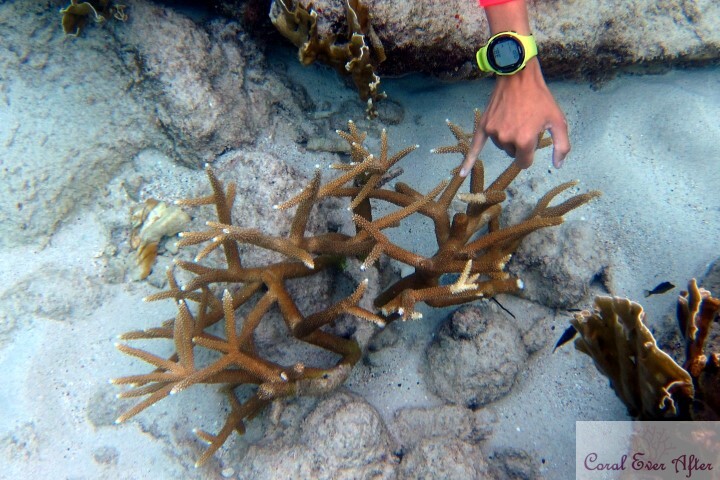
A gall crab‘s home is circled in red (small tube toward the top of the coral). This is an example of a growth anomaly, where the coral grew around the gall crab’s tube. Although gall crabs are commonly associated with dying coral, the gall crabs are likely not a sufficiently substantial environmental stressor to be the root cause.

Here’s a little troublemaker Blenny (ok, but in all fairness, it’s pretty cute). Notice the growth anomaly, where the coral’s skeleton is growing down into the Blenny’s home.

Coral with Growth Anomaly

Similarly, this maze brain type coral has a lovely structural pattern surrounding a creature’s home (I did not stick around long enough while scuba diving to determine the inhabitant). The structure is interesting to me – the coral terminates many times down at the hole. I wonder if this provides the coral some type of benefit – maybe it functions as a food distribution source (i.e., the inhabitant’s leftover’s/waste become the coral’s next meal).

Back to top

Lesion Relief:

Lesion Reliefs are other Growth Anomalies and include umbonate lesions (rounded growth), exophtic lesions (growing out beyond the surface), nodular lesions (small mass), and bosselated lesions (small bits that protrude). An example of exophtic lesions is below. While I do have other photographic examples of lesion reliefs, they may not be clear enough to distinguish between the other three lesion relief types.

This staghorn Acropora coral was covered in these growth anomalies. During my quick swim past while scuba diving, I was unable to discern the cause. The growths (exophtic lesions) appear like the skeleton has broken through the tissue and then become covered with cyanobacteria.

Growth Anomalies of Unknown Cause (GAN):

Growth Anomalies of Unknown Cause include various areas of accelerated growth, chaotic polyp arrangement, “gigantism,” and other as yet undetermined causes of unusual growth.

It appears some algae may have colonized prior areas of damage on this coral and created growth anomalies.

Back to top

Tissue Loss Lesions:

Various coral predators (e.g., flatworms, fish, sea stars, other corals), infections (e.g., viral, bacterial, fungal), mechanical interference (e.g., falling rockwork, directed water flow, sedimentation), chemical interference (e.g., high alkalinity with low nutrients), and as-yet unexplained issues may all cause tissue loss. The tissue loss lesion appearance may provide clues to the root cause, such as blenny bite marks shown previously.

Dying Orange Fungia Plate Coral

Another coral stung this Fungia coral, which resulted in a tissue loss lesion. Although the damage was severe, the coral did survive (click here to learn more).

Back to top

Other Soft Tissue Lesions:

Catalaphyllia Elegance Coral Syndrome

“Elegance Coral Syndrome” (ECS) is what I’d consider an outlier for gross tissue lesion types. The initial signs are external mesenterial filaments with retracted polyps and stubby tentacles. This isn’t a tissue pigmentation issue, tissue loss, or a growth anomaly (at least to my non-medically trained brain). Do we need a new category for soft tissue changes beyond simply its presence/absence and color?

Back to top

Skeletal Lesions:

Skeletal anomalies do not fall within the tissue response categories (according to some sources, but other sources include them), but it is a gross change to the coral that can result in one or more of the lesion types above, especially tissue loss or growth anomalies. I’ve included them here for completeness.

Poorly Fragmented Euphyllia Coral

Skeletal damage from poor fragmentation, damaging rockwork (or storms, if in the ocean), fish bites, and other root causes can result in lesions. This Euphyllia coral was poorly fragged, which resulted in a tissue loss lesion and ultimately death.

On the left is a slice of a Euphyllia coral skeleton that was infested with a boring sponge, while the Euphyllia coral skeleton slice on the right was not affected. Notice the dissolved areas on the left. While this type of damage is not a tissue lesion, it may result in lesions.

Back to top


Coral Disease Assessment – Lesion Location:

Where the lesion is located on a coral can provide clues toward the coral disease assessment for a root cause. For example, certain bacteria are sediment-associated, and sediment typically aggregates along the base of the coral. As another example, some fish are corallivorous, and they eat while swimming, which means the top of the coral is typically the most damaged. Therefore, the lesion locations are broken up into basal/peripheral (base or around the perimeter), medial/central (middle/center), and apical (top). Of course, severe or extreme cases could present as combination locations.

Basal/Peripheral Lesion Location:

Basal or peripheral lesions are ones that originate at the base or outer perimeter of the coral. As pictured, this coral has a lesion that originated at its outer perimeter along the base. (I found this coral while scuba diving, and I do not know its long-term outcome.)

Back to top

Medial/Central Lesion Location:

Coral Disease Assessment:  Medial and Multifocal Lesions from Predation

Medial or central lesions occur around the middle of the coral. The coral colony pictured does have a few lesions that would fall under the “apical” category, as they are along the top of the coral, but hopefully this is still a sufficient example of centrally-located lesions. (I found this coral while scuba diving, and I do not know its long-term outcome.)

Back to top

Apical Lesion Location:

Coral Disease Assessment:  Apical Tissue Loss from Fireworm Predation

Apical lesions are found at the top of the coral. The Acropora coral pictured has a few branches with lesions at the tips, caused by fireworms. (I found this coral while scuba diving, and I do not know its long-term outcome.)

Back to top

Combination Lesion Locations:

There are lesions covering these coral colonies, for a combination presentation (more common in severe or extreme cases of lesion severity). The lesions are along the base, at the top, and all across the center. (I found this coral while scuba diving, and I do not know its long-term outcome.)

Back to top


Coral Disease Assessment – Lesion Size

Documenting the lesion size is, in theory, fairly straightforward, as it is just the measurement of affected tissue. However, coral structure, obstructions, and indistinct margins can complicate the measurement. And, of course, the measurement units must be consistent in documentation (e.g., using centimeters or inches).

Back to top


Coral Disease Assessment – Lesion Duration:

Simply observing the coral’s skeleton can provide a good indication of how long the lesion has existed, and, if monitored over time, how fast the lesion is progressing. White skeleton indicates recent necrosis (about a few days), while a light green skeleton may indicate necrosis of a week or more ago. Skeletons populated with multiple algal types indicate necrosis in the more distant past (possibly months or more).

Acute Lesion Duration:

Dying Lobophyllia

The left-center of this coral shows an Acute Lesion Duration, with the white skeleton showing no algal colonization. Notice the far-left side of the coral skeleton, where there is a film algal colonization along with filamentous algal growth.

Back to top

Subacute Lesion Duration:

Dying Lobophyllia

No, you aren’t seeing double (at least not unintentionally). The far-left side of the coral’s skeleton represents a Subacute Lesion Duration, where filamentous algae is beginning to colonize on the coral. This is in contrast to the left-center of the coral, where there is an Acute Lesion Duration showing the white skeleton.

Back to top

Chronic Lesion Duration:

Dying Lobophyllia

Although there is a portion of an Acute Lesion Duration to the center-left of the coral shown with white skeleton, the majority of the remaining coral skeleton shown represents a Chronic Lesion Duration. There is a gradation of algal colonization, with caulerpa, bubble algae, and coralline algae growing across the coral’s skeleton.

Back to top


Coral Disease Assessment – Lesion Severity:

Lesion severity is the percentage of affected coral and ranges from mild to extreme. Interestingly, I had a difficult time finding mild and moderate examples to show, as most of the corals I’ve rehabilitated fell into the severe or extreme categories. Typically, I do not attempt to salvage mild or moderately affected corals, as they tend to heal without any intervention.

Mild (<10%) Lesion Severity:

This Echinophyllia (chalice) coral had a mild lesion on one side, where the lesion size was less than 10% of the coral size. This is an example of a mild (<10%) lesion severity.

Back to top

Moderate (10-24%) Lesion Severity:

Scolymia Rehabilitation

This Homophyllia (“Scolymia”) coral had a moderate lesion (let’s assume this one is 24%, although it is close to bumping up into the “severe” lesion severity category) due to a coral sting. This is an example of a moderate (10-24%) lesion severity. To learn more about this coral and its recovery, click here!

Back to top

Severe (25-49%) Lesion Severity:

Back to top

Stung Wellsophyllia Coral

Similar to the Homophyllia coral, another coral severely stung this Trachyphyllia coral. Notice how the lesion was just shy of half of the coral. This is an example of a severe (25-49%) lesion severity. This was a severe case, but the coral made a full recovery. To learn more about this coral’s journey, click here!

Back to top

Extreme (50%-100%) Lesion Severity:

Water-Deteriorated Lobophyllia Coral

This Lobophyllia coral had multiple issues, with lesions covering more than half of the colony. Despite such a bad condition, this coral recovered in only four months. This is an example of extreme (50-100%) lesion severity. Read more on this coral here!

Back to top


Coral Disease Assessment – Lesion Color:

Lesion colors are important to coral disease assessment, as they may indicate potential disease to consider (e.g., white patch disease). Lesion colors are highly interesting to me, and I haven’t found enough research to support what I’ve been seeing with my corals. I’ll add more here as I learn more.

Purple Lesion Color:

Coral Disease Assessment: Dark Spot Syndrome with purple lesions

The lesions depicted are purple (and are a combination of annular/irregular with a coalescing distribution). This was the first example I had seen of purple lesions aside from Aspergillosis fungus in Gorgonian corals and may be an example of Dark Spot Disease (DSD). (I found this coral while scuba diving, and I do not know its long-term outcome.)

Back to top

White Lesion Color:

Wellsophyllia Dying

Although these lesions are not entirely white, white lesions are areas of bleached tissue. This coral had two large areas of bleached tissue, but it made a full recovery. To learn more about this coral’s recovery, click here!

Back to top

Pink Lesion Color:

Dying Favia

Distinguishing a pink lesion from coralline algae may be a bit tricky, but in this case, the pink coloration is possibly originating as a defense mechanism from the coral. Despite the coral’s poor condition, it made a full recovery. Click here to learn more!

Back to top

Blue Lesion Color:

Coral Disease Assessment:  Pigmentation Lesion

This coral has both blue and white lesions; the blue coloration is not natural, as it occurred post-bleaching during recovery. In my experience, sometimes corals with bleached lesions will heal back with blue coloration. However, these corals rarely survive long-term after the initial recovery, in my experience (my guess is that the blue coloration is from hosting a particular zooxanthallae population that is insufficient to sustain the host coral – it is almost like they become non-photosynthetic corals – or at least insufficiently photosynthetic). This is such a recurring challenge for me that I call it “Bleached-Blue-Death” given the progression from bleaching to blue coloration to death. When I have rescued corals like this, I continue spot feeding them long-term. This seems to work so far as a way to keep them alive. (I found this coral while scuba diving, and I do not know its long-term outcome.)

Back to top

Yellow Lesion Color:

Aspergillosis lesions (yellow with a purple band) on a Gorgonia coral

This Gorgonia coral has Aspergillosis, which starts with deep purple lesions that can expand and develop yellow centers.


Coral Disease Assessment – Lesion Pattern:

Lesion patterns, especially when combined with other lesion characteristics, can help provide a holistic picture for coral disease assessment. These patterns help determine if the cause may be bacterial, due to corallivore predation, or just due to poor water quality.

Annular Pattern:

This isn’t the best example of an annular lesion pattern, but it’s the best I have. An annular lesion is shaped like a ring, with healthy tissue typically on both sides of the ring. Unfortunately (and surprisingly), this coral did not recover.

Back to top

Irregular Pattern:

Dying Homophyllia Bowerbanki Coral

An irregular shaped lesion has edges without any sort of pattern or form. The lesion edges are not smooth or straight. Although this Homophyllia had multifocal and coalescing lesions, it still survived and was one of my favorite rescues. Click here to learn how all this decay still recovered!

Back to top

Linear Pattern:

Linear lesion shapes are in a straight line (or straight but curving along the skeleton in rounded or massive bouldering corals). This Micromussa (Acanthastrea) coral likely had “Brown Jelly Syndrome” (the coral is pictured in a dip, with the brown jelly-like substance already removed); this damage entirely occurred overnight. Thankfully, I was able to stop the infection and salvage the coral.

Back to top

Circular Pattern:

Similar to the “annular” pattern, both are round-shaped. However, the “annular” shape is more of a ring-shaped lesion with a healthy center, whereas the “circular” lesion pattern is entirely the lesion.

Oblong Pattern:

The “oblong” pattern is similar to the “circular” pattern, in that the entire lesion is in an oblong shape, rather than a circle. Crazy, I know. I’m sure I have a photo for this, but I feel like it wouldn’t add anything that your imagination hasn’t already addressed.


Coral Disease Assessment – Lesion Distribution:

How the lesion presents across the coral can also provide key insights to the coral disease assessment process. If the lesion is across the entire coral, then it could be a waterborne issue; whereas, a focal lesion could indicate physical interference or corallivore predation.

Diffuse Lesion Distribution:

Lithophyllon Coral in Quarantine

Diffuse lesions are spread across the coral, without a central location. This lesion presented as tissue decay across the highest points of the coral, with only tissue in the lower points surviving. Despite the coral’s terrible condition, it still fully recovered. Click here to learn more!

Back to top

Focal Lesion Distribution:

Focal lesions are very defined, with healthy tissue surrounding a central point. Initially, I thought something fell on the coral to cause the damage (e.g., a heater); however, I found flatworms were consuming it. Removal of the flatworms and the damaged portions resulted in the coral’s recovery.

Back to top

Coalescing Lesion Distribution:

Although this is likely a combination presentation with diffuse and coalescing lesions, it is still a good example of lesions across the coral growing and merging together – coalescing into larger lesions. Only a very small piece of this coral survived.

Back to top

Linear Distribution:

Coral Disease Assessment:  Distinct Linear Basal Lesion

Linear lesion shapes and linear lesion distributions are practically synonymous. The lesion progresses in a line across the coral (even if it is a bit of a curved line). (I found this coral while scuba diving, and I do not know its long-term outcome.)

Back to top

Multifocal Lesion Distribution:

Oh, this little fellow is a troublemaker for the host coral. Notice all of the white lesions around the hole (and the hole is a growth anomaly, as well). These lesions are from fish bites, so they are multifocal (more than one location). Although multiple fish bites in a central location could form a coalescing distribution appearance, a multifocal lesion description is more appropriate given that the lesions themselves are not growing.

Back to top


Coral Disease Assessment – Lesion Edges/Border:

Personally, I tend to mix up the lesion edges/border with the lesion margin/band. The lesion edge/border is the entire length of the lesion across the coral. This length should be much longer than the lesion margin/band, which is the distance from the apparent healthy coral to the lesion or coral skeleton.

Lesion edges (or the border of the lesion) are typically considered distinct/discrete or indistinct/diffuse, as shown below, to support in coral disease assessment. There is also a category for “annular,” but given that it’s already a lesion shape, I don’t see the value in having it describe the border as well. (I also don’t have a photo for an example, so maybe I’m just already biased against it.)

Distinct or Discrete Lesion Edges/Border:

Dying Cyphastrea Coral

The sharp contrast between the brown tissue and the white skeleton creates a distinct lesion edge (notice how some polyps are split between half-healthy and half-skeleton). Although this Cyphastrea coral only had a small portion of healthy tissue remaining, it recovered and eventually grew to one of the largest corals in my aquarium. Click here for the before and after photos!

Back to top

Indistinct or Diffuse Lesion Edges/Border:

Dying Cyphastrea Coral

Contrasted with the previous Cyphastrea with distinct lesion edges, the indistinct lesion edges on this Cyphastrea create difficulty distinguishing healthy tissue and the remaining skeleton. Thankfully, this coral made a full recovery. Read more about its journey here!

Back to top

Coral Disease Assessment – Lesion Margin/Band Type:

Lesion margins are the lines separating healthy tissue from unhealthy tissue or the coral’s skeleton. These are also frequently called “bands.” As previously mentioned, I tend to mix up the lesion margin/band with the lesion edge/border. The lesion edge/border is the entire length of the lesion across the coral. This length should be much longer than the lesion margin/band.

Smooth Margin/Band:

As previously shown, this lesion progresses in a smooth line across the coral (even if it is a bit of a curved line). (I found this coral while scuba diving, and I do not know its long-term outcome.)

Back to top

Serrated (or Irregular) Margin/Band:

This isn’t the best example of a serrated lesion, but when I think of serrated lesions, I think of coral predation, like corallivorous starfish or Acropora-eating flatworms. This poor Acropora was dying from a bit of everything but did make a full recovery.

Back to top

Serpiginous (or Irregular) Margin/Band:

A serpiginous lesion edge reminds me of a snake (i.e., serpent), where the edge winds tightly about the coral. Again, this coral made a full recovery.

Back to top

Undulating (or Irregular) Margin/Band:

Coral Disease Assessment: Acute Undulating Basal Lesion

Similar to the serpiginous lesion margins, the undulating lesion margins wind about the coral but in a less tight manner. (I encountered this coral while scuba diving, so I do not know its outcome.)

Back to top


Coral Disease Assessment – Margin/Band Thickness:

The lesion margin/band size is the measurement of the edge tissue, which, due to the narrow shape, may be difficult to obtain. And, in indistinct or diffuse lesions, the margin/band may be impossible to discern. Once again, the measurement units must be consistent in documentation (e.g., using centimeters or inches).

Back to top

Coral Disease Assessment – Margin/Band Color:

Lesion margin/band colors vary and provide good indicators for coral disease assessment. In fact, several named coral diseases reference the band color in the name – it is the primary indicator of the disease. Some of these include Black Band Disease, Yellow Band Disease, Red Band Disease, and White Band. Conversely, other syndromes and diseases have no distinct colored band, such as the Caribbean white syndromes. While I wish I had my own photos of these diseases to include here for educational purposes, I’m thankful that I have not yet encountered many obvious examples.

Coral with purple band lesion

This coral has a purple margin or band color, surrounding the lesion in the center. (I photographed this coral while scuba diving and do not know the outcome of the colony.)

Coral Disease Assessment:  Staghorn Acropora with Zip-tie Bands to Mark Disease Progression

This staghorn Acropora coral may have “White Band Disease.” Notice the zip tie placement near these bands toward the base of the coral. This physical indicator helps reef managers track the lesion growth and determine its growth rate. Also, there is a damselfish hiding in the top center of the photo. Damselfish nest in these corals and create algal beds. (I photographed this coral while scuba diving and do not know the outcome of the colony.)

Conclusion:

We’ve only begun to scratch the surface of coral diseases and how they may present, especially as Western Atlantic and Indo-Pacific creatures cohabitate within our aquariums. As you find new information on coral disease assessment, please reach out or share in the comments below. For additional information, please consider the following references.

References:

  1. “Coral Disease Handbook: Guidelines for Assessment, Monitoring & Management,” Global Environment Facility, June 2008. Website
  2. Hawthorn, Aine, et al. “An introduction to lesions and histology of Scleractinian corals.” Veterinary Pathology, vol. 60, no. 5, 31 July 2023, pp. 529–546. Website
  3. “Lesion Terminology and Visualization.” Coral Disease & Health Consortium, 13 Nov. 2023. Website
  4. Edited by Cheryl M. Woodley et al., Diseases of Coral | Wiley Online Books, Wiley Online Library.

Rehabilitation Journey of a Dying Dipsastraea Coral (Moon Coral)

Rehabilitation Journey of a Dying Dipsastraea Coral (Moon Coral)

When I first started keeping a saltwater aquarium, I was looking everywhere for inexpensive live rock (college debt was all-consuming).  Down in the rubble bin of a local fish store, I happened across a neat semi-spherical piece.  The owner asked $5 for it, which looking back was quite expensive for a 0.5lb piece of rock, but oh well. Little did I know that I would have a beautiful piece originating from a previously dying moon coral.

Dying Dipsastraea Coral (Moon Coral)

When I got the piece of “live rock” home, I realized it had about the most pathetic coral remaining on it.  I tried to not get my hopes up, and I definitely didn’t see any beauty potential in it. When I placed the rock in the aquarium, the crabs swarmed over it like bees on honey.  There apparently was a fair bit of algae on it.  And, since this was 2007, there was no quarantine, no dipping, or any other good practice.

Please don’t mind the poor quality photo. This was at some point in 2007 (likely around July/August). I lost the original version somewhere along the way to 2024 (all that remains is a tiny enlarged thumbnail), my camera wasn’t the best, and admittedly, my photography skills were never great from the start.

Feeding a Dipsastraea Coral (Moon Coral)

 
After a few weeks, it started to extended tentacles, so I fed it. I was new to feeding corals, so it was probably just eating some pellets.

In this embarrassing photo (likely taken around December 2007), please don’t mind the bleached Alveopora and the fake colored blue ridge coral. I learned my lessons.

Recovering Dipsastraea Coral (Moon Coral)

The coral changed very little from ~July to December 2007 (~5 months), so the change from December 2007 to April 2008 (4 months) still amazes me.  During that time I upgraded from power compacts to metal halides, and my system reached over 1 year maturity. (Just look at that coralline!)

A

Recovering Dipsastraea Coral (Moon Coral)

After one year (~July 2008), the coral was not only healthy, but it was also becoming quite beautiful! And, my reefkeeping skills were a lot less terrifyingly bad!

Recovering Dipsastraea Coral (Moon Coral)

Note the incredible size difference between this photo (~October 2008) and the previous (~July 2008). The coral appeared to double in only about three months.

Rehabilitated Dipsastraea Coral (Moon Coral)

By late October 2008, there was almost no evidence the coral ever had any issues.

June 2009 (the latest photo I can find has this watermark)
 
I sold the coral in 2011 as it had fully outgrown my aquarium space, and we had to move across the country.  If I remember correctly, the final diameter was about 8″.  I never imagined my little piece of “live rock” would turn into such a beauty.
Dying Dipsastraea Coral (Moon Coral)Rehabilitated Dipsastraea Coral (Moon Coral)

Rehabilitation Journey of Dying Ricordea Corals

Previously Dying Ricordea Corallimorph Fully Rehabilitated

When I saw a rock at my local fish store with two dying Ricordea corals bleached to practically zero color, I knew I had to try to rehabilitate them.  

Intake: Acclimation, Inspection, and Dips

Bleached, Retracted,  and Dying Ricordea Corallimorph

Upon arriving at my home, I worked through my typical inspection, dip, and quarantine processes on these two dying Ricordea corals. Although hard to tell, this retracted Ricordea coral was nearly colorless (bleached) (March 14, 2012), but there was fluorescence. The mouths weren’t gaping, and the corals were intact (no damaged tissue). The bleaching severity led me to react to these corals as an “urgent” situation. The “usable diagnosis” (I’m not a medical professional, but this helps me create a care plan) was bleaching due to high light. (I saw where these two corals were physically located in the aquarium, so it was pretty obvious that they were receiving excessive light.)

Then, I had to assess the corals’ overall health. Since bleaching is often due to excessive light, wherein the zooxanthallae leave the coral, the coral has an increased chance of starvation. Note that the coral is surrounded by colorful coralline algae, which can typically tolerate higher-light conditions. This particular Ricordea coral was stressed but appeared in relatively good health (not quite starving). However, the other Ricordea (not pictured – I can’t find a photo) was extremely small and appeared on the brink of starvation.

Next, I removed the dying Ricordea corals from the substrate as much as possible to reduce introduction of parasites and nuisance algae. I continued through my dipping process, which included both a hydrogen peroxide-based dip and a CoralRx dip. Afterward, I glued each coral to a frag plug and placed both corals in the quarantine aquarium.

Quarantine: Care and Feeding

Although “soft corals,” like these dying Ricordea corals, can have a higher saturation point (level of light for optimal photosynthesis) and possibly photoinhibition point (level of detrimental light) than their stoney counterparts, they can still get stressed and expel their zooxanthallae.  With proper water conditions and lower light, they will frequently heal on their own.  Supplemental feeding helps replace the energy source previously provided by the zooxanthallae.

Recovering Ricordea Coral

Therefore, the solution for these corals was a healthy tank, low lighting, low flow, and a bit of feeding.  Ricordea are not the most apt to taking food directly, especially when unwell.  But, within a few days, the larger coral was eating well (very very small particulate food.) Once the corals went through 30 days of quarantine, were eating well, and did not have any concerning indicators, I used the dip process again before moving the Ricordea corals into my grow-out tank.

(Coral shown on 13 May 2012)

Bleached and Dying Ricordea Coral

The second Ricordea was not recovering as fast as the larger one, but it was starting to regain color.

(Coral shown 14 May 2012)

Recovering Ricordea Coral

By 22 May 2012, the larger Ricordea coral had almost fully recovered. The smaller one still had a ways to go.

Conclusion: Ricordea Rehabilitation Success

Previously Dying Ricordea Corallimorph Fully Rehabilitated

By 30 June 2012 (only slightly more than three months later), the larger Ricordea was fully rehabilitated. The smaller Ricordea made a full recovery as well.

Bleached, Retracted, and Dying Ricordea CorallimorphPreviously Dying Ricordea Corallimorph Fully Rehabilitated
From bleached to beautiful in only three months!

References:

For more information on light saturation and photoinhibition in corals and clams, see this link. (It’s a bit dated, but it still contains some interesting information.)

Dying Homophyllia (Scolymia) Coral Rehabilitation

Previously Dying Homophyllia australia (Scolymia) Coral Fully Rehabilitated

A local reefkeeper contacted me about possibly saving his dying Scolymia coral (Homophyllia australis), and the condition of it sounded quite bad. But, seeing its condition was still a shock. I wasn’t sure if there was even a mouth left since the tissue was so shrunken and discolored. But, I went ahead and did my best; its complete rehabilitation was a success! Read on for more information about this dying Homophyllia australis (Scolymia) coral’s successful rehabilitation.

Intake: Acclimation, Inspection, and Dips

Dying Homophyllia australia (Scolymia) Coral in a Dip

When I brought the coral home (12 March 2022), I placed it (still in the container) into the sump of my quarantine tank to acclimate to the temperature for only 15 minutes. Next, I brought it over to my workbench for inspection, dipping, etc.

First, I had to assess its health. For more information on assessing a coral’s health and potential causes of damage, check out “Assessing Coral Health: An Introduction.”

Next, came the inspection and dip process. My “handy-dandy flow-chart” led me to classify this coral as an “urgent” case, which meant that it was in bad shape requiring lots of care. However, it wasn’t to the point of an emergency. This category determination was based on the algae-covered skeleton, clinging tissue, and unknown mouth situation (along with the mesenterial filaments coming out everywhere). Then came my dip process, which was fairly standard for this case. Typically I would remove excess skeleton and glue down any damaged tissue, but in this case, I absolutely had no clue what was what. Even what little tissue remained didn’t fluoresce much, so I just rolled with what I had.

Quarantine: Care and Feeding

Dying Homophyllia australia (Scolymia) Coral

After the dips, I placed the coral into my quarantine tank. Typically, I place these types of corals on the sand bed, but I wanted to really observe this one closely. So, I placed it up on a frag rack up near the surface of the water. This would also make feedings easier. I still wasn’t sure what was tissue and what was skeleton at this point.

In several days (18 March 2022), I was able to start to see the actual remaining tissue condition a bit better. Granted, it was pretty bad. At this point, I was in a “watch-and-see” mode. Attempts to feed could cause the coral to rot, if it is unable to properly digest the food. However, I did start target feeding corals nearby to see if I could elicit a feeding response.

Dying Homophyllia australia (Scolymia) Coral

In less than a month (8 April 2022), the coral was no longer retracted into the skeleton. It wasn’t pretty, but by this point, I had confidence in its survival – as long as I could get it to eat. However, I still wasn’t ready to start feeding it, as its mouth condition was unsuitable.

Recovering Homophyllia australia (Scolymia) Coral

The coral was finally ready to start taking supplemental food by about the 1.5 month point (21 May 2022). Notice the tentacles extending and mostly-formed mouth. Success!

Conclusion: Scolymia Rehabilitation Success

Rehabilitated Homophyllia australia (Scolymia) Coral

Today, the previously dying Homophyllia australis (Scolymia) coral is an absolute stunner (although my photography is lacking). Notice how a lot of the grayish stripes turned green over time. This coral also features various orange shades and even yellow hues! Absolutely lovely! (Photo was taken 25 November 2023.)

Dying Homophyllia australia (Scolymia) Coral in a DipRehabilitated Homophyllia australia (Scolymia) Coral

Fossilized Oceans: Beautiful Agatized Coral Reefs

Indonesian Agatized Coral Cabochon

With this post, CoralEverAfter expands from just coral conservation of present-day corals to appreciation of their fossil history – and maybe a glimpse into the future. I’m a nerd, through and through, right down to the rock collection I’ve had since I was a kid.  However, last year, everything changed.  My husband went to Australia and brought me back some opals – in the rough.  He knew I had my Gryphon band saw for cutting live corals, so he thought the hard part was over (this reminds me of when I found that *free* fish tank at a yard sale that started my reefkeeping obsession).  Well, I ended up with a slab saw (to cut the rough into something more workable), and then I ended up with a cabbing machine.  And, of course, I couldn’t start with the opals; I had to practice!  So I started buying rocks…and got addicted.  But then, I found fossilized oceans.  More specifically, agatized corals that were once part of an ancient reef. 

So now, not only am I interested in reefkeeping and scuba diving, I’m also into coral lapidary arts.  But, it doesn’t stop there.  Ooooh no… Because what’s the point of having all these pretty rocks just stored up??  So, I started metalsmithing copper and silver.  I already bead, so now I have pretty much the whole lifecycle of jewelry. I hope you enjoy the amazing variety of coral polyp structures, mineral colors, cyanobacteria structures, preserved deathbeds of clams, and other finds on these ancient coral reefs from fossilized oceans.

Indonesian Coral necklace, set in copper with pearl and copper accents

Indonesia Agatized Coral and Marine Life

The variety of patterns and colors in Indonesian agatized coral and other marine life is truly stunning. Some are nearly entirely agatized, leaving little traces of the original flower-patterned mineralization. Others, leave just the polyp structure, resulting in an incredible field-of-flowers pattern. But, one of my absolute favorites isn’t even a fossilized coral; it’s “Script Jasper,” which is a mortality plate of clams made of vibrant oranges, puce, and small crystal patches.

  • Indonesian Agatized Coral Cabochon
  • Indonesia Agatized Coral and Rough
  • Indonesian Agatized Coral Cabochon and Rough
  • Indonesian Agatized Coral Cabochon
  • Indonesia Agatized Coral Cabochon and Rough
  • Coquina Clam Mortality Plate Cabochon and Rough
  • Indonesian Agatized Coral Slab
  • Indonesian Agatized Coral Slab
  • Indonesian Agatized Coral Slab
  • Indonesian Agatized Coral Slab
  • Indonesian Agatized Coral Slab
  • Indonesian Agatized Coral Slab
  • Indonesian Agatized Coral Slab

United States Agatized Coral and Marine Life

Southwest United States:

While most Utah agatized coral comes in varying shades of browns, grays, and yellows, the Red Horn Coral (Lophophyllidium sp.) stands out, not only amongst Utah corals, but is probably at the top worldwide for its coloration, patterns, and specimen quality. Another red fossilized coral is found in Arizona. Arizona fossilized coral is a unique combination of red jasper with a soft yellowish calcite center and bits of sparkly crystal druzy. While they may not make the highest quality cabochons for jewelry, they are certainly interesting in structure and coloration. But then, there are the Michigan corals. They deserve their own paragraph.

Midwestern United States:

Probably the most infamous fossilized coral is the Michigan Petosky Stone, known for its gray body and darker gray eyes. But, there are so many more corals overshadowed by the Petosky. Take, for instance, the lowly chain coral (Halycites sp.). What a fascinating coral structure that looks like a chain necklace! Then, there are Charlevoix stones, Cladopora, and so many more! Although I don’t have examples here, other parts of the Midwest are home to lovely fossil corals.

Southern United States:

While the Petosky stone probably holds the most popular title, Florida’s Tampa Bay agatized corals are downright breathtaking. However, if you’re a coral purist, you may find yourself saddened that very few of the specimens display any remnants of the coral polyps. These agatized corals are so fully agatized that very few traces of the original structure remain. In the slideshow below, the orange piece shows miniscule traces of the original polyps along the white outer edge.

Northwestern United States

If all the fossilized corals in the United States weren’t enough, there’s other fossilized sea life, particularly Stromatolites and Oncolites, which are various microbial remnants, primarily mats and orbs of cyanobacteria. And… let’s not forget the famous Turritella agate (although it’s a freshwater snail species).

  • Utah Agatized Horn Coral Cabochon and Rough
  • Utah Horn Coral Cabochon and Rough
  • Arizona Agatized Coral Cabochon and Rough
  • Michigan Chain Coral ("Halycites") Cabochon and Rough
  • Michigan Petosky Stone Cabochon and Rough
  • Tampa Bay, Florida Agatized Coral Cabochon
  • Wyoming Crinoid (Sea Lily) Cabochon and Rough
  • Stromatolite (Cyanobacteria Mat) Cabochon and Rough
  • Oncolites (Cyanobacteria orbs) Cabochon and Rough
  • Utah Horn Coral Slab
  • Michigan Cladopora Coral Slab
  • Michigan Petosky (Hexagonaria percarinata) Slab

Morocco Agatized Coral and Marine Life

It’s incredible to me that one of the most dazzlingly yellow pieces here is stromatolite (microbial mat, primarily cyanobacteria). The ketchup-and-mustard combination, swirled together amidst crystallized valleys, creates a stunning piece. In contrast, while the Actinocyanthus coral cabochon may not boast spectacular coloration, it compensates with exquisite detail in the polyp structure. Some specimens simply look like modern specimens filled with resin; they’re incredible.

  • Morocco Stromatolite (Cyanobacteria Mats) Cabochon
  • Morocco Actinocyanthus Cabochon and Rough

Mexico Agatized/Crystallized Coral

I rarely find agatized coral from Mexico (in fact, I only have two slabs), but both are extraordinary. They feature botryoidal formations (bubbly-round structures), crystals, and agate mixed amongst the jasper in varying shades of pinks, grays, and browns.

  • Mexico Agatized/Crystallized Coral Cabochon
  • Mexican Agatized Coral Rough
  • Mexican Agatized and Crystallized Coral Rough

United Kingdom Agatized Coral

My only specimen from the United Kingdom is what I believe is this Lithostrotion coral. It features crystalline tubes surrounded by a dark jasper. No polyp structures are evident.

  • United Kingdom Lithostrotion Agatized Coral Cabochon and Rough

Unidentified Locale Agatized Coral and Marine Life

These are all unique pieces obtained from various sources, but they also came without any origin information. If you have any insights to these pieces, please let me know!

  • Unidentified Agatized Coral Cabochon and Rough
  • Invertebrate Mortality Plate Cabochon and Rough
  • Stromatolite (Cyanobacteria Mat) Cabochon
  • Unidentified Agatized Coral Cabochon and Rough

Conclusion:

Agatized corals are a great way to learn about ancient fossilized oceans and the predecessors to our current coral reefs. By studying the past, embracing the present, and planning for the future, maybe we can continue the fairytale of CoralEverAfter with a happy ending.

To Learn More:

If you would like to learn more about fossilized corals, their identification, and their structures, this website *rocks*!

Introduction to Pipe Organ Corals

Naturally white Pipe Organ (Tubipora musica?) coral

Pipe Organ corals (Tubipora musica) are absolutely amazing, in my opinion.  They build red tubes for each polyp, and the varying heights of those tubes makes the coral look like… wait for it… a pipe organ!  Their polyps extend out from the tubes and wave in the current. The green variety makes a great alternative to green star polyps, for those who like the wavy green mats but would prefer something a bit less invasive. With years of success keeping and sharing these unique beauties, hopefully this introduction to Pipe Organ corals will help others.

Pipe Organ (Tubipora musica) coral colony

When I entered the reefkeeping hobby back in the 00’s (wow – I’m old), Pipe Organ corals were on the no-go list, as they typically slowly diminished in most aquariums.  Of course, as a newbie, I didn’t do my research and bought a fantastic green one.  Somehow, some way, I managed to keep it… and it thrived!  Over the many years I had it, I fragged it over a hundred times.  This is it a bit browned out from high nutrients (note the green hair algae).

Naturally white Pipe Organ (Tubipora musica?) coral

Then, when a local fish store heard of my success, they gave me a dying white one (not bleached) to try.  Again, I had success! It turned into one of the most stunning corals I’ve ever had. (And, to be clear, the coral stayed a very slight peach color for years, as shown in this photo.)

So, based on my success over years, here is what I’ve learned. First, knowing what they are is key. Pipe organ corals are usually mistakenly categorized as “large polyp stony corals” as they form solid tubes, but they are actually classified as soft corals.  However, their care level is more intensive than most soft corals. 

Quarantine and Treatment of Pipe Organ Corals:

Recovering Pipe Organ (Tubipora musica) coral

Since the Pipe Organ coral is made of tubes, there are tons of perfect living spaces for all sorts of creatures.  Be aware that the pipe organ could harbor bristle worms, flat worms, and lots of detritus.  Use of a good coral dip and quarantine will help prevent unwanted organisms.

I took this coral in to rescue it. Note the complete lack of tissue on the tubes near the polyps. However, the skeleton is mostly clear of algae, and the coral’s degradation has occurred over a long period of time. If the skeleton was covered in algae, the coral would likely struggle to recover more. [Learn more about assessing coral health]

Lighting:

The green variety definitely requires higher light than the white variety, as it easily will brown out in lower light.  I usually kept the green ones up on the rocks with my SPS corals and the white ones down on the sand bed. Although, as with all corals, an initial acclimation period is recommended.

Flow:

Pipe Organ Coral (Tubipora musica) polyps

This is tricky.  Flow must be high enough to prevent detritus from settling on/in the tubes, but low enough to not whip the delicate polyps around.  Detritus settlement is a huge concern as it will fuel algal growth, which will, in turn, outgrow the coral.

Nutrients:

As stated above, nutrients are a huge concern, as any algae covering the Pipe Organ coral tubes will prevent the coral from extending its polyps.  I do not recommend pipe organ corals for aquariums having an issue with cyanobacteria, hair algae, or other invasive alga.  I’ve seen recommendations that Pipe Organ corals need pristine water parameters, with phosphate and nitrate levels toward those recommended for SPS (nearly undetectable). However, that has not been my experience. They seem to tolerate high nutrients well, as long as there is no nuisance algae on the colony.

In my experience, Pipe Organ corals also don’t appreciate target feeding.  I’ve tried various foods and various methods, but they all result in the coral polyps “wilting.”  As soon as the food touches their polyps, the polyps just shrivel and droop, which more resembles a defense mechanism than feeding.

Since the coral is made of tubes, there are tons of areas for detritus collection. If the coral is not opening, the tubes can be blown off lightly with a turkey baster to help remove detritus. The can tubes grow in a structure similar to the inside of bamboo with internal closures. If the coral is not kept free of detritus, portions of the tubes can trap the detritus indefinitely. This may leach nutrients from the colony over time and may cause issues in the future.

Other Water Parameters:

While I was not concerned with higher phosphate and nitrate levels for Pipe Organ corals, I was concerned with maintaining proper alkalinity, calcium, and magnesium levels. These corals may not be growing the typical SPS or LPS skeletons, but they are still creating massive structures. While I won’t debate the “proper” ranges here, I have always maintained my tanks around Alk: 9 dKh, Ca: 425 ppm, and Mag: 1350 ppm. But, that’s just me, and it seems to work just fine.

Fragging Pipe Organ Corals:

Pipe Organ (Tubipora musica) coral frags

I believe the lack of success from decades ago came from poor fragging.  Most colonies imported were chopped very close to the top of the polyp, which ended up chopping the body of the polyp itself.  I highly discourage the use of bone cutters on pipe organs since the cutters are more likely to crush the coral than cut it.  Instead, I recommend a razor blade.  Cut a healthy specimen from the top downward, hopefully going between tubes.  Keep as much tube height as possible as it is hard to determine where the polyp is inside.  Use cyanoacrylate (super glue gel) to mount the frag on a piece of rubble or plug.  Within about a week, small fleshy bumps should appear, as shown, as the start of new tubes. They usually start about a centimeter down from the top of the red tube and form a horizontal base around the tube. It is these horizontal attachment points that the razor blade should cut through during fragging, rather than the tubes themselves.

When All Else Fails:

If a pipe organ dies, its skeleton is still useful.  They are gorgeous additions to fish-only aquariums due to their bright red natural coloration.  They are also great in sumps due to their porosity (great hiding places for all sorts of critters).  My favorite way to recycle the skeleton is to crush it up and add it to a white sand bed.  The red dots contrast beautifully with a stark white background.

Conclusion:

Pipe Organ corals are a beautiful addition to a reef tank, and they do not deserve the reputation they have received. Their red skeletons provide a bright pop of unique color, and their polyps provide interesting movement within the tank.       

Pregnancy and Reefkeeping

Pregnancy and Reefkeeping

[If you are experiencing a medical emergency, please contact 911 or whatever emergency service is available in your area!]

In honor of Mother’s Day this year in the United States, this post covers the considerations and risks of keeping a saltwater aquarium while becoming a
mother. Many years ago, I was pregnant with my first child, and I was appalled at the little amount of information available on pregnancy and reefkeeping. Unfortunately, as I’m reposting this article, there is still very little information. If you’re reading this post thinking, “I’m not pregnant, so I don’t care,” then at least read it for the safety of those around you. A lot of the medical risks exist regardless of pregnancy status. Hopefully my experience and referenced scientific studies will help someone! If you prefer to avoid the scares, you can skip to the bottom for recommendations.

Disclaimer: I am not a medical-related professional in any capacity. If you have a medical concern, seek help from a professional. This article is provided only for basic information and awareness.

Changes in Smell during Pregnancy

An abnormal sense of smell was reported in 76% of pregnant women in a study published by Oxford University Press.1 In early pregnancy, I was continuously nauseous. The smell of “low tide” was practically unbearable and only worsened my nausea. Water changes were particularly gut-wrenching. Later in pregnancy, the smell did not bother me as much.

Ergonomic Considerations

Water weighs approximately eight pounds per gallon (or roughly a kilogram per liter for the rest of the world). Lifting water for water changes or top-off may be excessive, depending on the health/ physical condition of the woman. I was used to carrying five gallons of water for top-off every couple of days, but later in pregnancy I was unable to safely lift that amount of weight. Talk to your doctor about how much weight is safe for you to lift.

My tank was a “tall” tank at 31″ high in addition to the 30″ stand. In order to reach into the tank, I had to use a step-stool. Medical professionals sometimes frown upon pregnant women elevating themselves due to the risk of falling. Although a small step-stool may seem like little-to-no risk, I had a non-pregnant friend fall and break her wrist while she cleaned her tank. Pregnant women may find themselves with less balance than usual due to a change in their
center of gravity and loosened ligaments.

Potential Pathogens in Reefkeeping that can Affect Pregnancy

Here’s the real problem. The scientific community knows very little about marine environments, especially how they interact with humans (let alone pregnant humans). The following information is not meant to scare anyone; it is meant to educate. As many pregnant women will attest, there is enough scary information out there (eat this, but don’t eat that; work out, but not too much; be calm, but worry about everything!) There is absolutely no way to
avoid all risks. I educated myself the best I could, and then I made the decision to continue maintaining my aquariums. My favorite part of reefkeeping is rescuing dying/diseased animals, and due to that elevated risk, I decided to stop rescuing while I was pregnant. In my opinion, there are too many known pathogens that are transmissible to humans in order to safely treat diseases while pregnant. Considering how much the scientific/medical community learns every day about pathogens, I did not want to be the first case of some bizarre new disease while pregnant.

Vibrio

For those of you who follow my rescuing endeavors, you probably know how much I hate Vibrio. It’s a gram-negative bacteria that is incredibly difficult to treat in marine animals. It often presents itself in humans as sepsis (septicemia), gastrointestinal distress, and/or open sores. This bacteria is one of the
reasons that pregnant women are told to not eat raw seafood. Some of the antibiotics most effective against Vibrio may not be safe for pregnant women, so a less-effective medication may have to be used. I use Kanamycin on my fish, and this antibiotic is known to cause hearing loss in a developing fetus. Your doctor will have to weigh the medication benefits/risks against the severity of the infection.2

Brucella

This is another gram-negative bacteria present in marine environments and is usually associated with eating raw seafood. It can cause granulomas in the brain, and it may present with symptoms similar to meningitis.3 Again, the most effective antibiotics may not be safe for pregnant women.2

Mycobacterium

This bacteria is most commonly associated with Tuberculosis, and it is transmissible from marine animals to humans. As an example, sea lions at a zoo in the Netherlands were infected with Mycobacterium, and six of their 25 handlers tested positive for the disease.4 There have been several documented cases of Mycobacterium marinum infections in reefkeepers (not pregnant), and you can see their disturbing images here.5 Additionally, the medications used to treat Tuberculosis can cross the placenta to the fetus.6

Aeromonas

The phrase, “Don’t drink the water when travelling abroad,” is often due to Aeromonas spp. bacteria. It is usually associated with gastrointestinal distress.7 Once again, the drugs required to treat the illness are not entirely safe with pregnancy.

Erysipelothrix

This is a gram-positive bacteria usually associated with animal-centric occupations (e.g., veterinarian, biologist, farmer). Usually it presents as a cutaneous lesion, and it can lead to complications.8

Others

There are many other bacteria associated with marine environments and animals known to cause disease/illness. Some of these include Edwardsiella, Salmonella, E. coli, etc.9

Bites, Stings, Poison, & Venom:

Unfortunately, this category is too large to discuss in great detail. Many simple coral stings may be easily treatable with vinegar followed by hot water, but an eel bite usually requires medical treatment with antibiotics and/or stitches. Knowing the difference between poison and venom is an important start to the discussion. Poison is defined as “a substance that through its chemical action usually kills, injures, or impairs an organism,” according to the Merriam-Webster Dictionary.10 Similarly, the dictionary defines venom as, “poisonous matter normally secreted by some animals (as snakes, scorpions, or bees) and transmitted to prey or an enemy chiefly by biting or stinging”.11 Therefore, venom is essentially poison used intentionally to cause damage.

Palytoxin

Palytoxin dangers are highly debated, but regardless of the nuances, it is still one of the most toxic poisons known. It has a “lethal dose (LD59) in mice of 0.15 microgram per kilogram by intravenous injection”.12 It is most commonly associated with palythoas (zoanthids), but it is also associated with dinoflagellates, crabs, fish, etc. (another reason why pregnant women should not eat raw seafood).13, 14, 15 There have been several human fatalities due to palytoxin (usually through ingestion of contaminated seafood). Further research determined, “its intravenous LD50 in the dog, rabbit, monkey, guinea pig, rat, and mouse range between 0.033 and 0.45 μg/kg.” This same study determined palytoxin is relatively non-toxic when administered intragastrically. It is an irritant and can cause tissue damage when applied topically.16 While most of us wouldn’t dream of injecting ourselves with palytoxic intentionally, many of us have been poked significantly with vermetid worm tubes or other sharp spines. Palytoxin is still quite toxic subcutaneously and dermally (so don’t rub a zoanthid on
yourself, get tank water in your eyes, or place an open wound into your tank).

Corals:

Corals contain nematocysts (stinging cells) on their tentacles that inject through human skin like tiny barbs. The sting from a coral may be painless, feel like an electric shock, itch like poison ivy, or even cause anaphylactic shock. For instance, fire corals inject a protein venom that causes a burning sensation (hence the name, “fire coral”).17

Other Animals:

Eels, lionfish, rabbitfish, urchins, and many other animals are known to bite, sting, or otherwise puncture. Sometimes, even the most innocuous creature will turn for the worst. I had a bicolor blenny who loved to bite me. Any open wounds, especially when caused by a marine creature, are subject to infection. Know what creatures you have in the tank that could create a wound or envenomate.

Chemicals

Many chemical products have a “Material Safety Data Sheet” (MSDS), or as it is now called, a “Safety Data Sheet” (SDS). These sheets list the ingredients, known hazards, and basic first aid information. Unfortunately, many of our aquarium products state, “proprietary blend”. Although unspecified ingredients are still required to have hazard information, there is still some risk due to unknowns (someone might be particularly sensitive to an unspecified ingredient). Pregnant women should particularly pay attention to the sections on “Teratogenic Effects” and “Developmental Effects”. These parts states what is known to cause harm to a developing baby. Since most women are unwilling to have invasive experimentation performed on their babies, there is very little information on the risks of various chemicals during pregnancy. You can find many of the common MSDS (SDS) through a simple search.

Consider looking up the following chemicals, if you use them. It is best to know the chemical manufacturer during the search, but that is certainly not always possible. I looked these all up back in 2015, and this is the information I found at that time (it may not be accurate today, but this is an example of what results may be.)

  • Calcium chloride – anhydrous (used to make calcium supplements): Teratogenic effects are not available.
  • Calcium hydroxide (kalkwasser; used to raise calcium and alkalinity): Teratogenic effects are not available.
  • Magnesium chloride (used to make magnesium supplements): Teratogenic effects are not available.
  • Magnesium sulfate (used to make magnesium supplements): Teratogenic effects are not available.
  • Sodium bicarbonate (used to make alkalinity supplements): Teratogenic effects are not available.
  • Sodium carbonate (used to make alkalinity supplements): Teratogenic effects are not available.
  • CoralRx (coral dip): Teratogenic effects are not available.
  • Bayer Advanced Insecticide (sometimes used as a coral dip): “DEVELOPMENTAL TOXICITY: In an oral developmental toxicity study in rats treated with beta-cyfluthrin, decreased fetal body weights and an increased incidence of skeletal findings were observed at the maternally toxic and lethal high dose level (40 mg/kg).”
  • Levamisole (used as a coral dip): “DEVELOPMENTAL TOXICITY: Classified Reproductive system/toxin/female [POSSIBLE].”
  • Lugol’s Iodine (used as a coral dip): Teratogenic effects are not available.
  • Red Sea Reef Foundation Test Kit (tests calcium, alkalinity, and magnesium): “Mg Reagent A contains Sodium tetraborate decahydrate, but at levels below the threshold for classification. This compound has been found to have effects on reproduction and fertility. No compounds present in the reagents have been identified as having carcinogenic, mutagenic properties.”

Recommendations for a Pregnancy and Reefkeeping:

You can significantly reduce your risks to the pregnancy while reefkeeping by avoiding aquarium maintenance. However, if you decide to continue caring for aquariums, then I recommend speaking with a medical professional, considering the following the subsequent advice, and continuously re-evaluating your risk tolerance. Be honest and up-front with your doctor, and take the provided medical advice seriously. Wear gloves and other personal protective equipment as much as possible. Less exposed skin is better.

If you are fragging, then also wear eye protection (a face mask and face shield are advisable as well). Limit lifting, climbing, or other strenuous activity, especially if your doctor recommends against it. After aquarium maintenance, wash every exposed body part with hot water (as hot as is safe) and soap. Scrub thoroughly. Do not touch your face before you wash. Also, please do not eat, drink, or smoke while performing aquarium maintenance (or smoke at all while pregnant). This will help limit ingestion of poisons/bacteria.

Be familiar in first aid, and have those in your household also become familiar in first aid. For any injuries, contact medical personnel. Typical treatment for most aquarium stings/bites/etc. is to soak the area in vinegar for about 15 minutes, followed by a soak in hot water (as hot as you can safely withstand without burns) for about 30-90 minutes. The vinegar will help dissolve the nematocysts and prevent them from further discharging. Many poisons/venoms are broken down by heat (although some are intensified with heat, so contact medical personnel!)18 If you develop any rashes, lesions, or develop odd symptoms (loss of vision, heart palpitations, wheezing, numbness, etc.), then also contact medical personnel immediately!

Stay up to date on your immunizations (particularly tetanus if you plan to continue reefkeeping). Make a list of your aquarium inhabitants and potential exposures, and provide this to your doctor. I also kept a list of these things along with other relevant medical information in my purse. Sometimes medical issues will surface long after the initial exposure, so it’s important to have the information written in case you are unable to relay potential exposure information. After the baby is born, make sure the aquarium area is baby-proofed. Curious little hands will find every hazard!

Conclusion on Pregnancy and Reefkeeping:

Although I continued to perform aquarium maintenance, I stopped rescuing coral/fish due to the increased risks. I experienced no aquarium-related injuries and had a healthy baby. Although this is not a comprehensive list of marine hazards to a developing baby and its mother, hopefully it is an educational start. Reduce the risks where possible, and if you must accept some risk, be prepared on how to handle it. Good luck! If you can think of other hazards, let me know in the comments!

References:

Note: These references are now quite dated; the links may not work and/or the information may be superceded. Please review the latest studies and speak with your medical professional.

  1. Nordin, Steven, et al. “A Longitudinal Descriptive Study of Self-reported Abnormal Smell and Taste Perception in Pregnant Women.” Chemical Senses (2004) 29 (5): 391–402.
  2. Norwitz ER, Greenberg JA. “Antibiotics in Pregnancy: Are They Safe?” Reviews in Obstetrics and Gynecology. 2009;2(3):135-136.
  3. Sohn AH, Probert WS, Glaser CA, et al. “Human Neurobrucellosis with Intracerebral Granuloma Caused by a Marine Mammal Brucella spp.” Emerging Infectious Diseases. 2003;9(4):485-488.
  4. Hu, G, et al. “Meta- and Pooled Analyses of the Effect of Glutathione S-transferase M1 and T1 Deficiency on Chronic Obstructive Pulmonary Disease.” The International Journal of Tuberculosis and Lung Disease 12, no 12, December 2008, 1474-1481.
  5. Pro, Steven. “Mycobacterium marinum: The Fish Disease You Could Catch”, Reefkeeping, 2003, http://www.reefkeeping.com/issues/2003-07/sp/feature/.
  6. Centers for Disease Control and Prevention, http://www.cdc.gov/tb/publications/factsheets/specpop/pregnancy.htm, accessed 2 May 2015.
  7. Vila J, Ruiz J, Gallardo F, Vargas M, Soler L, Figueras MJ, et al. “Aeromonas spp. and traveler’s diarrhea: clinical features and antimicrobial resistance.” Emerg Infect Dis [serial
    online] 2003 May [date cited].
  8. Reboli, A C, and W E Farrar. “Erysipelothrix Rhusiopathiae: An Occupational Pathogen.” Clinical Microbiology Reviews 2.4 (1989): 354–359. Print.
  9. Clarridge, J E et al. “Extraintestinal Human Infection Caused by Edwardsiella Tarda.” Journal of Clinical Microbiology 11.5 (1980): 511–514. Print.
  10. “Poison,” http://www.merriam-webster.com/dictionary/poison, accessed 10 May 2015.
  11. “Venom,” http://www.merriam-webster.com/dictionary/venom, accessed 10 May 2015.
  12. Richard E. Moore and Paul J. Scheuer, “Palytoxin: A New Marine Toxin from a Coelenterate,” Science, 30 April 1971: 172 (3982), 495–498.
  13. Arthur M. Kodama, Yoshitsugi Hokama, Takeshi Yasumoto, Masakazu Fukui, Sally Jo Manea, Neal Sutherland, “Clinical and laboratory findings implicating palytoxin as cause of ciguatera poisoning due to Decapterus macrosoma (mackerel),” Toxicon, Volume 27, Issue 9, 1989, Pages 1051-1053.
  14. Angel C. Alcala, Lawton C. Alcala, John S. Garth, Daisuke Yasumura, Takeshi Yasumoto, “Human fatality due to ingestion of the crab Demania reynaudii that contained a palytoxin-like toxin,” Toxicon, Volume 26, Issue 1, 1988, Pages 105-107.
  15. Yutaka Onuma, Masayuki Satake, Takanori Ukena, Jean Roux, Suzanne Chanteau, Noelson Rasolofonirina, Mamy Ratsimaloto, Hideo Naoki, Takeshi Yasumoto, “Identification of putative palytoxin as the cause of clupeotoxism,” Toxicon, Volume 37, Issue 1, January 1999, Pages 55-65.
  16. J.S. Wiles, J.A. Vick, M.K. Christensen, “Toxicological evaluation of palytoxin in several animal species,” Toxicon, Volume 12, Issue 4, August 1974, Pages 427-433.
  17. Eric Borneman, “Venomous Corals: The Fire Corals,” Reefkeeping, November 2002. http://reefkeeping.com/issues/2002-11/eb/
  18. “Marine Animal Bites or Stings,” http://www.nlm.nih.gov/medlineplus/ency/article/000032.htm, accessed 10 May 2015.

Breeding Journal, Snail Species: Columbellid euplica

Collumbellid snail, also known as Hawaiian Strombus

Given its aliases as “Hawaiian Strombus” and “Strombus maculatus,” the Collumbellid euplica snail may not have its nomenclature, especially to the species level, all settled. But that doesn’t change how awesome of a snail it is. As shown below, they reproduce faster than rabbits (or for that matter, faster than my Stomatella hitchhiker snails!) They stay small, graze over rocks, and fit into the smallest nooks and crannies. As the algal population waxes and wanes, so, too, does their population for a harmonized tank. Back when Collumbellid snails were covering my tank, I decided to join the Marine Breeding Initiative. This was my journal entry documenting the breeding. Some of it may be a bit repetitive, but it was required for the breeding documentation.

Collumbellid snail, also known as Hawaiian Strombus
Collumbellid Snail, also known as Hawaiian Strombus Snail

General Information:

General Species: Columbellid euplica

Social Structure: Group

Size of Individuals: 1/2″ long

Age of Individuals: Unknown

Date added to Tank: May 2009

Collumbellid Snail, also known as Hawaiian Strombus Snail

Broodstock Tank Details:

Size of Tank: 40g
Substrate Details: Bare bottom
Filtration Details: Live Rock, Skimmer, Carbon/GFO Reactor
Water Changes: 10% Weekly
Water Temperature: 76 degrees
Lighting: 6 x 39W T-5
Lighting Cycle: 8 hours on
Other Tank Inhabitants: Blue legged crabs, nerites, corals

Broodstock Tank Feeding Details:

Food Types: Formula Two Pellets and DIY Frozen Blended Seafood
Feeding Schedule: 2x/week

Spawning Details:

Date of First Spawn: Within days after introduction to the aquarium
Spawn Time of Day: Unknown

Dates of Consecutive Spawns: Constantly?

Courtship Details: None apparent yet

Egg Size: <1mm

Egg Color: White

Egg Count: 6-7 in each packet

Collumbellid Snail Egg Masses laid on 1/27/2012
Collumbellid Snail Egg Masses laid on 1/27/2012 - closeup
Collumbellid Snail Egg Masses laid on 1/27/2012 – closeup
Collumbellid Snail Egg Masses status on 2/2/2012 - closeup
Collumbellid Snail Egg Masses status on 2/2/2012 – closeup
Collumbellid Snail Egg Masses status on 2/6/2012 - closeup
Collumbellid Snail Egg Masses status on 2/6/2012 – closeup

Hatch Details:

Hatch Date: 2/6/2012

Hatch Time of Day: Unknown

# Days after Spawn: ~28 days

Larvae Description: Adult-like

Hatching Collumbellid snails
Hatching Collumbellid snails (from a different spawn)

Larval Tank Details:

Temperature: 76
Size of Larval Tank: 40g
Substrate Details: Bare Bottom
Other Tank Decor: Live Rock
Filtration Details: Live Rock, Skimmer, GFO/Carbon Media Reactor
Lighting: 6 x 39W T-5
Lighting Cycle: 8 on
Water Changes: 10% weekly

Larval Tank Feeding Details:

Food Types: Algae
Feeding Schedule: Constant

Metamorphosis/Settlement:

Date of Settlement Start: N/A
Days after Hatch: N/A
Date of Settlement End: N/A
Description of Fry: Appear as small adults

Juvenile Collumbellid Snail Size Reference with Hydroid
Juvenile Collumbellid Snail
Week-old Juvenile Collumbellid Snail

Grow-Out Tank Details:

Temperature: 76
Size of Grow-Out Tank: 40g
Substrate Details: Bare Bottom
Other Tank Decor: Live Rock
Filtration Details: Live Rock, Skimmer, GFO/Carbon Media Reactor
Lighting: 6 x 39W T-5
Lighting Cycle: 8 hr on
Water Changes: 10% weekly
Size at Transfer: N/A
Age at Transfer: N/A

Grow-Out Feeding Details:

Food Types: Algae
Feeding Schedule: Constant

Additional Information:

Update 14 June 2012:  I’ve found that these snails are able to make some sort of mucus thread that they are able to slide down in order to drop themselves lower in the tank.  I also got to watch a snail glide across the top of the water surface, make a thread (only held by the surface tension), and then drop down to the bottom of the tank.  It’s quite fascinating to watch!

Great Reference Info (with photos):  http://www.advancedaquarist.com/2009/11/aafeature

error: Contact us to request digital rights.